-
PFPS
PFPS
Pain, stiffness, an injury that won’t heal — if it’s keeping you from moving normally, we likely treat it. Here are the conditions we see most across our 8 Texas clinics. No referral needed to start.
“Patellofemoral Pain Syndrome Explained: Signs, Symptoms, Risk Factors, Diagnosis, and Cutting-Edge Physical Therapy Approaches”
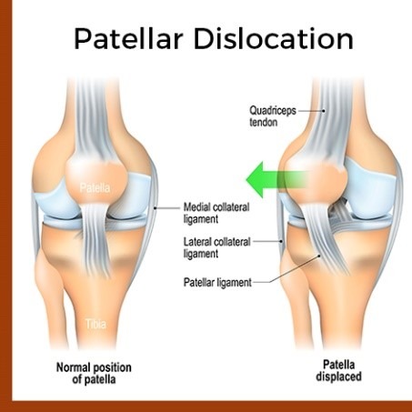
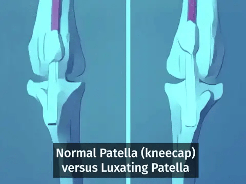
What is Patellofemoral Pain Syndrome (PFPS)?
Risk Factors:2,3
Muscular imbalance like quadriceps weakness or altered firing pattern or weakness of hip muscles.
Overuse or wrong training patterns.
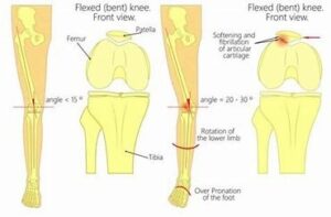
Malalignment or altered biomechanics of hip, knee or foot.
Patellar hypermobility or anomalies like patella alta or hypoplasia of medial patellar facet.
Tightness in iliotibial band or lateral retinaculum, quadriceps, hamstrings or glutues.
Trauma or previous surgeries.
Diagnosis and examination:2,3
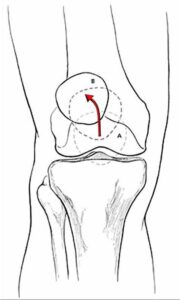
Lateral patellar tracking sign: Suggests patellar maltracking as a result of tight lateral restraints or VMO dysfunction. ( J sign)
Tenderness present in Medial or lateral retinaculum common in PFPS.
Poping or clicking may present Patella may produce a palpable pop, click, or clunk with palpation during passive or active range of motion; may be a sign of patellar maltracking, perhaps caused by synovial hypertrophy, plica synovialis, or cyst formation; a popping sensation with marked lateral deviation of the patella in extension indicates patellar instability.
Patellar glide: Assesses patellar mobility; displacement of less than one quadrant indicates tight lateral structures; displacement of more than three quadrants suggests patellar hypermobility caused by poor medial restraints.
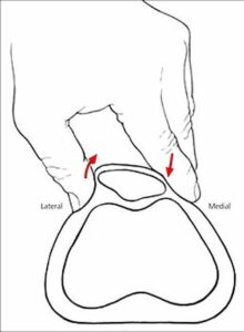
Patellar tilt test: lateral aspect of patella is fixed and cannot be raised to at least horizontal position indicates tight lateral structures.
Patellar apprehension: pain or discomfort with lateral translation of the patella indicates lateral patellar instability as a cause of pain.
Flexibility of quadriceps, hamstrings, ITB, hip flexors, and gastrocnemius can be assessed as compromised.
Gait and foot analysis for pronated foot or compensatory gait mechanics.
Treatment:
1. Exercise Therapy:4,5,6,7
Strengthening exercises: Isometric exercises while the knee is fully extended used at the beginning of the therapy, because it minimizes stress on the patellofemoral-joint. Straight-leg exercise, patient elevates extended leg and holds it for 10secs or pillow squeeze exercise Patient sits comfortable with his trunk supported. Both knees extended. Placing a pillow/towel under one knee Patient is asked to push the pillow/towel in the table by extending his or her knee. Later on which are progressed to closed and open chain exercises like Squats in pain free range or terminal knee extension. Strengthening of VMO and hip muscles are also added to off load the knee joint in activities.
Stretching of targeted muscles: Tightened iliotibial tract, hamstrings, gluteus and quadriceps stretching is given passively to give better results with long duration of hold to improve muscle flexibility.
Core Stabilization exercises: It enhances the overall stability and reduces knee stress, proper engaging of all core muscles is taught in rest as well as during exercises or activities.
2. Proprioceptive Training: 8,9
3. Biomechanical Assessment and Correction: 1,2,3
4. Manual therapy:
Techniques such as joint mobilizations, soft tissues release of tightened structures, and Myofasical release can be used to address muscle imbalances.